Mr Arthrogram Hip
Paralabral cysts in the hip joint:.
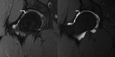
Mr arthrogram hip. An arthrogram is ordered to:. MR Arthrogram Wrist. An elusive source of hip pain case reports and literature review.
The exam is done in two parts and usually with the aid of a contrast agent called gadolinium that will help to highlight the visualization of joint structures and improve the MRI evaluation. It's simple, secure and free. Affordable cash prices allow everyone access to this important imaging modality, even patients who have no.
R/O TFCC, SL or LT Ligament Tear. 180 Providers in these locations. Relevant imaging should be reviewed, and the details of the patient confirmed.
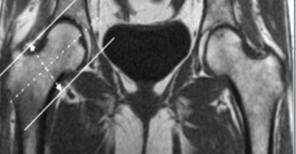
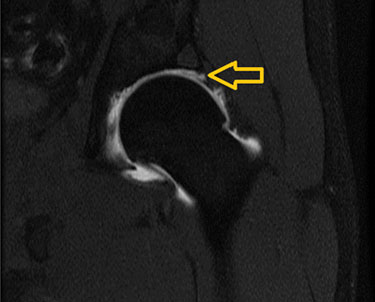
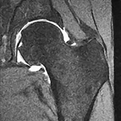
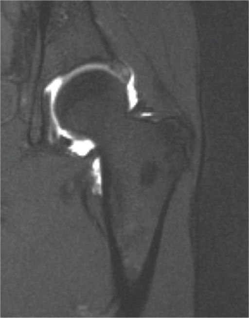
The upper image from an MR arthrogram of the right hip is normal. , &. It may be done alone or with other tests such as an MRI scan or CT scan.
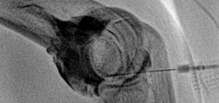
A needle is placed into the joint for the arthrogram and contrast and/or medication are put into the joint after taking out any fluid from the joint. Particularly when labral tears, femoroacetabular impingement, or. 24 Subsequent publications confirmed the advantages of MR arthrography in patients with chronic hip pain;.
Arthrography is commonly done on the joints of the shoulder and knee. Low-Cost Arthrograms with Detailed, Accurate Reporting An arthrogram is a complicated procedure, but that doesn’t mean you’ll have to pay a fortune for this common diagnostic imaging procedure. , &.
Arthrograms can be diagnostic and therapeutic. A 43-year-old male asked:. This procedure may also be performed on the wrist, elbow, ankle and hip.
In addition, in locations without access to an. A very small subchondral cyst was noted along the lateral roof of the right acetabulum What does this mean?. How do I prepare for an arthrogram?.
Ziegert AJ, Blankenbaker DG, De Smet AA, et al. 175 Providers in these locations 175 Providers in these locations. Arthrography is the x-ray examination of a joint that uses a special form of x-ray called fluoroscopy and a contrast material containing iodine.
Mr arthrogram At our PMI Diagnostic Imaging suite in New Lenox, we also offer MR Arthrograms. MR Arthrogram Ankle. MRI ARTHROGRAPHY (W/ CONTRAST ONLY) MRI (MAGNETIC RESONANCE IMAGING) CT ARTHROGRAPHY (W/ CONTRAST ONLY) Cardiac Stress Test (4 CPT codes required) multi study.
I had an MRI arthrogram on my right hip in 06. Read verified reviews from patients like you and see real-time availability for every doctor. In 1995 and was shown to better depict the acetabular labrum than conventional MR imaging.
Confirm an intra-articular position with imaging;. Your healthcare provider will tell you how to prepare. An arthrogram is a joint x-ray used to find the cause of pain or check healing after surgery.
MR Arthrogram Elbow. It may be done if standard X-rays do not show the needed details of the joint structure and function. In the hip – to show any tear of the cartilage labrum (or rim of the joint).
Ultrasound of Hand to Rule Out Nonradiopaque Foreign Body. MRI Hip Arthrogram CT Hip Arthrogram Knee Arthrogram X-ray Knee Arthrogram MRI Knee Arthrogram CT Knee Arthrogram Wrist Arthrogram X-ray Wrist Arthrogram MRI Wrist Arthrogrm 731 CT Wrist Arthrogram 3D Reconstruction. Hip Arthrogram D,E 93yoM ©Ken L Schreibman, PhD/MD 15 schreibman.info Example:.
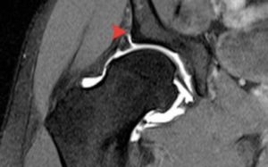
The upper right circle is of the scapholunate ligament, while the top left circle is the lunatotriquetral ligament. This will make the hip feel tight. The upper image from an MR arthrogram of the right hip is normal.
The arthrogram is often used to complement X-rays and radiographs. The hip is a medium-sized joint and the injected volume should reflect this. The injection itself can cause localized pressure and pain.
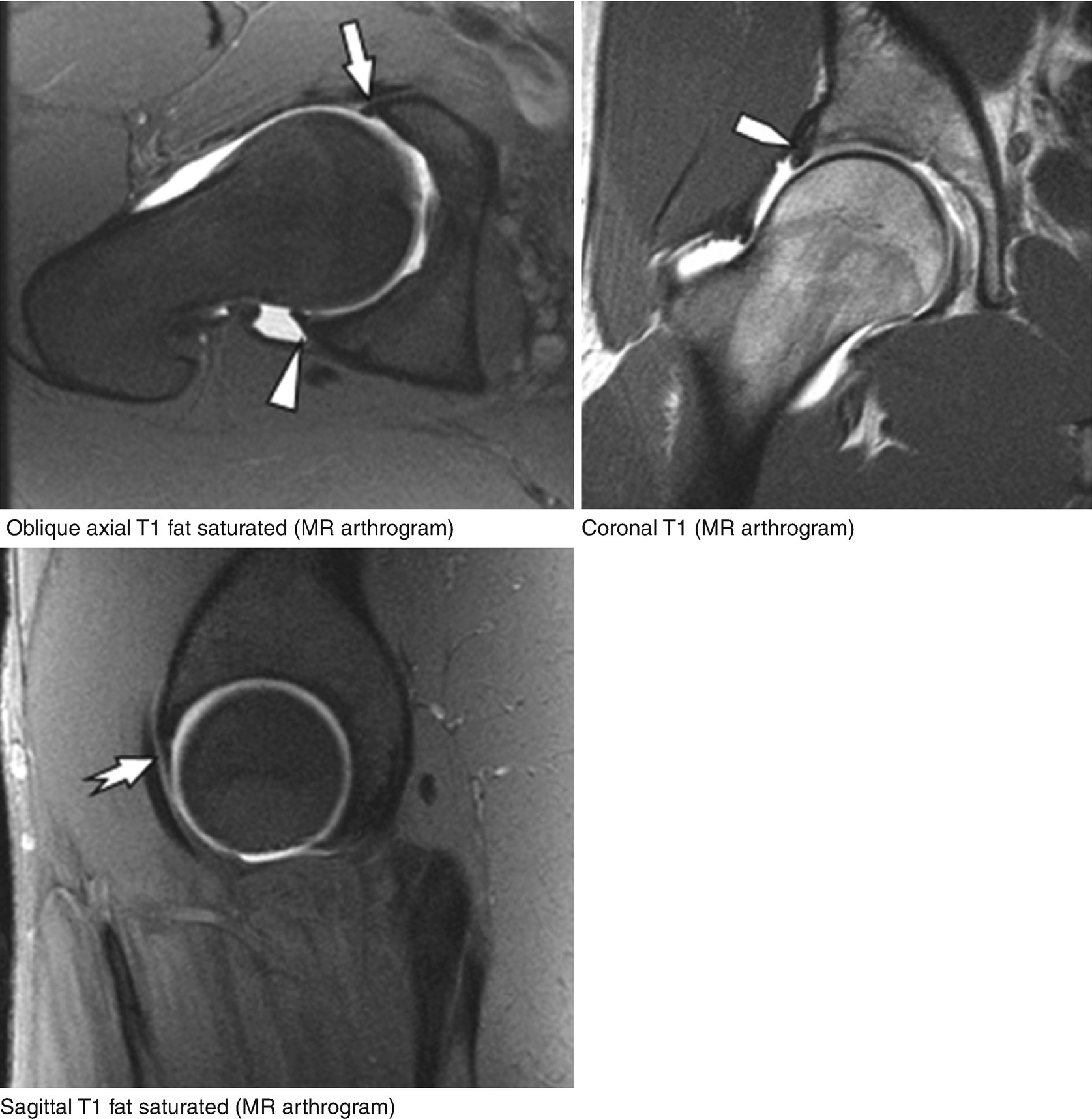
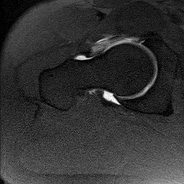
First, an Arthrogram will be performed. Detachments are more common than tears and are identified on the basis of the presence of contrast material interposed at the acetabular-labral junction. Arthrography requires an intra-articular injection of GBCA into the joint space.
It was the most excruciating pain, despite him topping up the anesthetic 3 times.(As someone who has lived with extreme chronic pain for half my life, and experienced countless medical interventions, I‘m no drama queen!) i knew instantly that something was VERY wrong. Some common reasons for an arthrogram are:. This page is for OHSU's MRI technologists and physicians.
The process is quite simple and includes having dye contrast injected into the joint and then undergoing MR imaging to get pictures of the joint. Our radiologists work closely with OHSU MRI technologists in the art of creating optimal images using current technology. Check that you're covered.
What is an MRI Arthrogram?. Book with doctors for an MRI arthrogram of the hip by using Zocdoc. A radiologist will inject contrast medium into the joint space for the evaluation of joint abnormalities.
Tutorial on performing a shoulder and hip arthrogram. Joint Injections and Aspirations. On MDsave, the cost of an MRI/CT with Arthrogram ranges from $1,040 to $2,773.
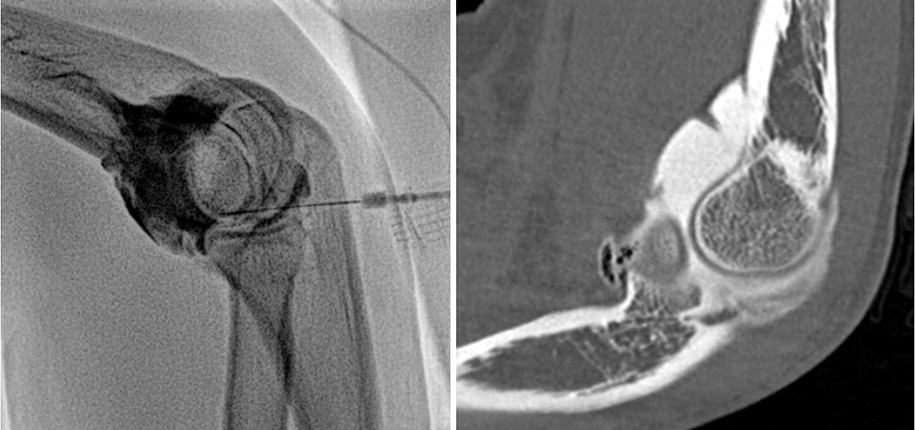
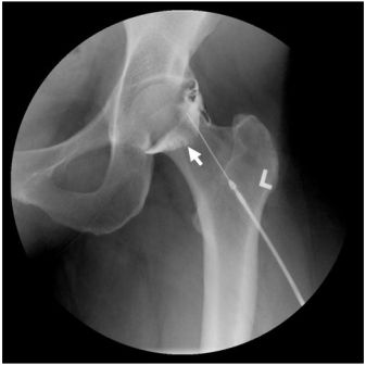
Occasionally, an MRI does not provide a full picture of certain structures in the body. You will receive a local anesthetic to numb to area. Once numb, contrast agents will be injected into your joint with the help of an image-guided device, typically a fluoroscope or CT scanner, to guide the injection into the right spot.
Read more about how MDsave works. Comparison of standard hip MR arthrographic imaging planes and sequences for detection of arthroscopically proven labral tear. Not as an "MRI or CT with contrast.".
MR ARTHROGRAM MRI CPT CODE/ FLUOROSCOPIC GUIDANCE CODE /INJECTION PROCEDURE CODE. Find and compare top local doctors. That’s when you might need an arthrogram, also called arthrography.
Tell healthcare providers if you are allergic to iodine or shellfish. A retrospective analysis of 113 patients who had MRI arthrogram and who underwent hip arthroscopy was included in the study. An arthrogram is a procedure that uses fluoroscopic-guidance (a special type of x-ray) to inject contrast dye directly into a specific joint.
Those on high deductible health plans or without insurance can shop, compare prices and save. , &. Injections for mri hip arthrograms are normally done under x ray fluoroscopic guidance (some radiologists prefer to use ultrasound).
MSK MRI Basic MSK MRI;. Rule Out Labral Tear. Pain Treatment and Therapy Program.
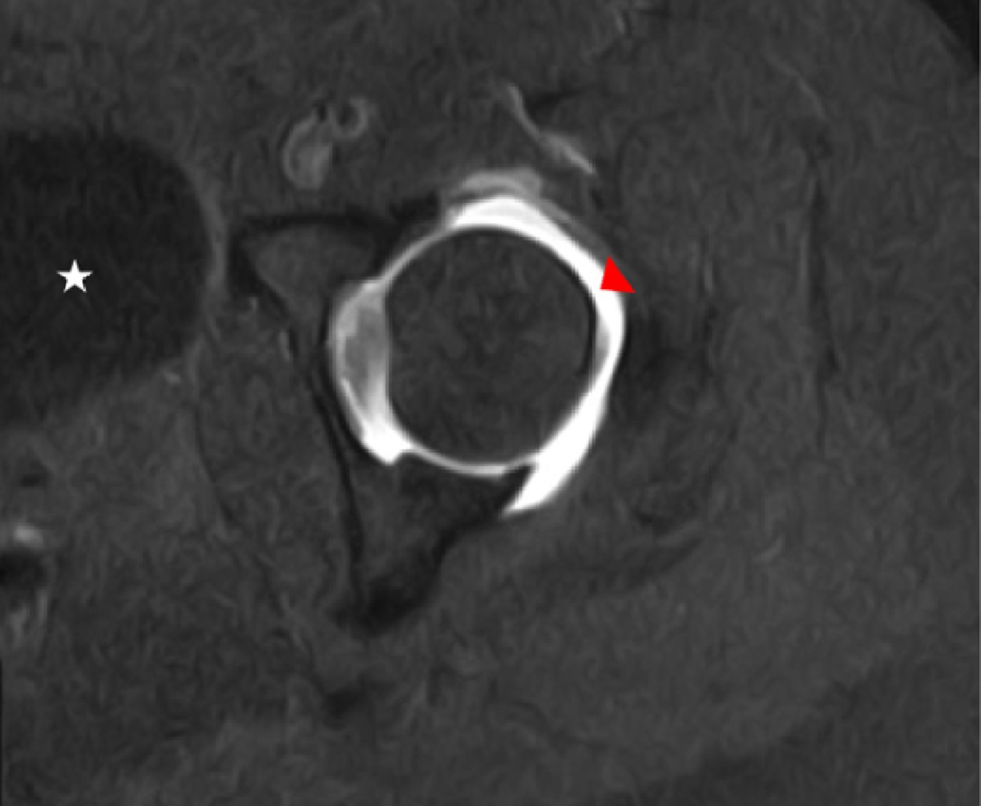
A pathologic labral condition, detachment or tear, is a common cause of chronic hip pain, and MR arthrography of the hip is the imaging procedure of choice for identifying an abnormal labrum. , & 242. Find a provider near you-Or-USE CURRENT LOCATION.
Findings at MR arthrography. An arthrogram is used to radiologically examine the tissues of the joints by injecting a contrast dye into the area. “We get a lot of additional information about the joint,” explains CDI Musculoskeletal Radiologist Dr.
The general principles of hip arthrogram injections are to:. Hip Arthrogram X-ray Hip Arthrogram MRI Hip Arthrogram CT Hip Arthrogram Fluoro Guided Knee Arthrogram & X-ray Knee Arthrogram & MRI Knee Arthrogram & CT Knee Arthrogram Fluoro Guided Wrist Arthrogram & X-ray Wrist Arthrogram. Your doctor may order an arthrogram when a standard X-ray is unable to show the source of pain or lack of movement in a certain joint, such as the hip, knee, shoulder or wrist.
AP of Pelvis **(5 v+ use CPT ) Dual-Energy X-Ray (DEXA). Joints, specifically shoulders, hips, knee, and occasionally wrists and ankles are unique because tears in tendons, cartilage, and ligaments are so small and covered by tissue that MRIs will often miss the injury site. In the wrist – to show any tear of the small ligaments of the wrist.
No preparation is needed. It’s another type of imaging where first you get a special dye, called contrast dye, injected into your joint. An MRI arthorgram is an imaging study conducted to diagnose an issue within a joint.
The same technique can be used for performing a therapeutic injection with anesthetics and steroids as. Arthrography is a type of imaging test used to look at a joint, such as the shoulder, knee, or hip. Dye is injected to help healthcare providers see your joint clearly.
If you have a prosthesis, you may need an arthrogram to see if it is loose. An arthrogram is an x-ray of a joint. Unlike a typical MRI, an MRI arthrogram begins with the injection of fluid called contrast right into the joint – usually a hip, shoulder, wrist, elbow or knee.
Do I need to do anything to prepare for the test?. An arthrogram is used to:. Therapeutic arthrograms often distend the joint with cortisone and lidocaine, with a common site being the shoulder.
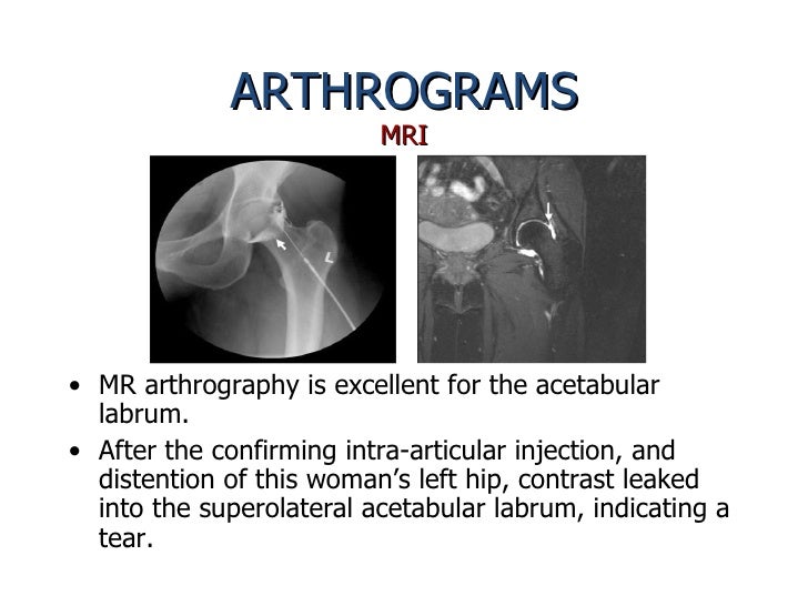
28 Magerkurth O, Jacobson JA, Girish G, et al. 18 years experience Medical Oncology. Evidence from a review of the radiologic literature supports the use of direct MR arthrography over unenhanced MRI and indirect MR arthrography for the detection of labral and cartilage abnormalities in the hip.
Magnetic resonance imaging (MRI) arthrography of the hip has been widely used now for diagnosis of articular pathology of the hip. An arthrogram is an interventional procedure for the assessment of joint capsule pathologies. , &.
If it is read as a cyst by a. It outlines all sequences and protocols currently applied in our MRI section. MR Arthrogram Shoulder.
MRI Arthrogram - Hip. AP of Pelvis DEXA (BONE DENSITOMETRY) Hip Bilateral, Incl. Intraarticular abnormalities of the hip are being diagnosed with increased frequency by diagnostic methods such as conventional MRI, conventional MR arthrography, and hip arthroscopy.
An MRI with injected contrast would be used to diagnose a possible tear in any of the three locations and should be ordered as an MRI arthrogram or, if MRI is contraindicated, a CT arthrogram;. MR arthrography is most often used in evaluation of the hip and acetabular labrum, of the shoulder rotator cuff and glenoid labrum, and less often in the wrist. In the shoulder – where the joint is unstable or if an ultrasound or plain MRI has not shown a suspected tendon tear.
Arthrography is an MRI technique used to image joints if a standard MRI does not provide enough detail. MR Arthrogram is a diagnostic imaging test used to evaluate your joint health, particularly in the shoulder, hip, and occasionally the wrist, elbow and ankle. The lower image demonstrates an unsuspected stress fracture of the femoral neck (arrow).
MRI ARTHROGRAM LEFT HIP ;. Use the Mouse to Scroll or the arrows. An MR Arthrogram is a two-part test.
A hip arthrogram is used to examine congenital abnormalities in the area. Hip Arthrogram D,E 93yoM ©Ken L Schreibman, PhD/MD 15 schreibman.info Skin Sub-Q Fat Keys to Arthrography ADVANCE UNTIL HIT BONE Don’t stop at the capsule WATCH FIRST DROP OF CONTRAST Should flow away from needle tip If contrast stays. MR Arthrogram Hip.
The hip is injected from an anterior approach 1,2. MR arthrography of the hip was first described by Hodler et al. Enjoy the videos and music you love, upload original content, and share it all with friends, family, and the world on YouTube.
Patient Safety Tips Prior to an Arthrogram Please let us know if you have any allergies or adverse. A 32-year-old woman complained of hip pain related to vigorous daily aerobics. An arthrogram uses imaging equipment to evaluate a joint like the shoulder, elbow, wrist, hip, knee or ankle.
The contrast material is then injected to distend the hip joint – iodinated contrast material if doing a conventional arthrogram or CT or dilute gadolinium, a heavy metal contrast material if being followed by MRI. The diagnostic accuracy of acetabular labral tears using magnetic resonance imaging and magnetic resonance arthrography:. An MRI is a very good test for bone and connective tissue.
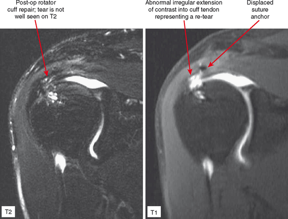
The most important indications for this technique over MR arthrography include a failed MR arthrogram, an obese or severely claustrophobic patient, a patient with an MR-incompatible implanted medical device, or a postoperative patient with metal hardware in close proximity to the joint ( Box 5-1 ). KNEE SHOULDER SHOULDER ARTHROGRAM ANKLE ELBOW WRIST HIP CONTACT. The more commonly performed MR arthrograms are of the shoulder and hip.
An arthrogram is a test using Contrast X-rays to obtain a series of pictures of a joint. Find tears, degeneration or disease in the cartilage, ligament or tendon. Images can show precise type and location of joint damage.
Rule Out Labral Tear. It is a two-part procedure consisting of a contrast injection into the joint, followed by an MRI or CT scan of the joint.
Q Tbn 3aand9gcryikapq1244og Japzrwcrltto3ivwa2i3zjxj4txmzlw9ipsk Usqp Cau
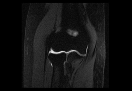
Arthrogram Mri Radiology Reference Article Radiopaedia Org

Mr Arthrography Versus Conventional Mri In Evaluation Of Labral And Chondral Lesions In Different Types Of Femoroacetabular Impingement Sciencedirect
Mr Arthrogram Hip のギャラリー

A 29 Year Old Female With 1 Year Of Progressive Hip Pain A Coronal T1 Download Scientific Diagram

Arthrogram Diagnostic Imaging Services

Mri Arthrogram Shoulder Rule Out Labral Tear Cedars Sinai

Coronal T1 Fat Saturated Mr Arthrogram Image Through The Midfemoral Download Scientific Diagram
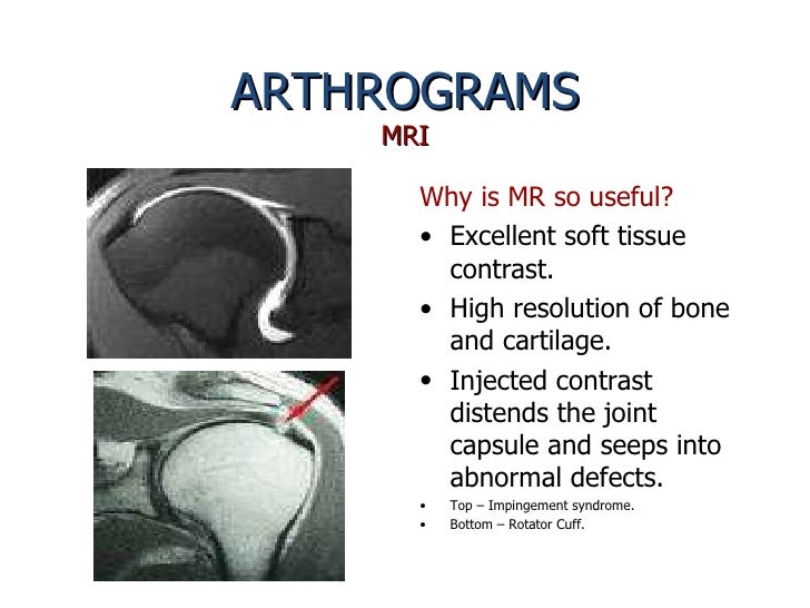
Arthrograms Presentation

Pdf Mr And Ct Arthrography Of The Hip Llopis Semantic Scholar

P 2 A Axial Hip Mr Arthrograms Diagram Quizlet
Mri Arthrogram Radtechonduty

Normal Hip Arthrogram Youtube

Hip Arthrogram Injection Fluoroscopic Guided Radiology Case Radiopaedia Org
Pelvis Hip Springerlink

Figure 5 From Imaging In Hip Arthroscopy For Femoroacetabular Impingement A Comprehensive Approach Semantic Scholar
Q Tbn 3aand9gcsdgrc0cbpf3kraqpeb26dtsg J0rnczsygu08hpvliespfgsxe Usqp Cau

46 Year Old Male With Hip Pain A T 2 Weighted Coronal Mr Image Download Scientific Diagram
Arthrogram For Mri Or Ct Cincinnati Children S Blog

Shoulder And Hip Mri Arthrogram Tutorial For Medical Personnel And Patients Youtube
Mr Hip Arthrogram Wo Msk Protocol Ohsu
Arthrogram For Mri Or Ct Cincinnati Children S Blog
Acetabular Labral Tear Radiology Case Radiopaedia Org

Mri Arthrogram Hip Protocol And Planning Indications For Mri Arthrogram Hip Scan

Imaging Of The Acetabular Labrum A Review Hard Tissue

Mri Vs Mra When Is An Arthrogram Needed Vista Health

Arthrogram Wikipedia

Mr Arthrography Proves Superior To Standard Mr In Labral Tears

Different Types Of Mris At Mbj Missoula Bone Joint

Coronal Magnetic Resonance Arthrogram Of The Hip The Bmj

Epos P 0027
Hip Labral Tears And Femoroacetabular Impingement A Frequent Cause Of Non Arthritic Hip Pain Derek Ochiai Md Nirschl Orthopaedic Center

What Do You Think About The Mr Arthrography Of The Hip
Cureus The Dark Side Of Gadolinium A Study Of Arthrographic Contrast At Extreme Concentrations

Mri Arthrogram
Mri Arthrogram Radtechonduty
Q Tbn 3aand9gcr9z7dfq4rsapxkujxq81x4zodo1ks7qea0j1yees Gs T6mfce Usqp Cau

Hip Labral Tear Knee Sports Orthobullets
Www Birpublications Org Doi Pdf 10 1259 Bjr
Arthrograms Presentation

Hip Mri
Journal Of Lancaster General Health Imaging Insights
Exam Details Peace Regional Mri Clinic In Dawson Creek
Labral Tears Gorav Datta

P 2 D Coronal Mr Arthrogram Hip Diagram Quizlet

Acetabular Labral Tears London Sports Orthopaedics

Xjlta Azwnioum
Http Pdf Posterng Netkey At Download Index Php Module Get Pdf By Id Poster Id

Mri Vs Mra When Is An Arthrogram Needed Vista Health

Chronic Adult Hip Pain Mr Arthrography Of The Hip Radiographics
Mr Hip Arthrogram Wo Msk Protocol Ohsu
Http Pdf Posterng Netkey At Download Index Php Module Get Pdf By Id Poster Id
Http Essr Org Content Essr Uploads 17 10 Ct Arthrography Mr Arthrograp Pdf
Q Tbn 3aand9gctakghfi50fjv0hxwdysr 0r1jqwu84svh1bwvx Edkqdcbrjx0 Usqp Cau

Normal Mr Arthrography Of The Hip Complete Mri Examination Youtube

Mri Arthrogram Hip Protocol And Planning Indications For Mri Arthrogram Hip Scan

Clicking Hip In A Postmenopausal Woman Cmaj

Epos Trade

Mr Arthrography Of The Hip With And Without Leg Traction Assessing The Diagnostic Performance In Detection Of Ligamentum Teres Lesions With Arthroscopic Correlation European Journal Of Radiology

Arthrograms Presentation

Acetabular Labral Tear Radsource

Mr Arthrogram Of The Right Hip Showing A Lesion Located At The Base Of Download Scientific Diagram

Diagnostic Imaging Of The Hip For Physical Therapists Physiopedia

Anterior Hip Review Intra Operative Imaging During Surgery

Mri Arthrogram Hip Rule Out Labral Tear Cedars Sinai
Http Www Jlgh Org Jlgh Media Journal Lgh Media Library Past issues Volume 4 issue 1 V4 I1 Wiggins Pdf
Mri Arthrogram Radtechonduty

Imaging Of A Painful Femoral Head Arthritis Research

Acetabular Labral Tear Radsource
Arthrography And Joint Injection And Aspiration Principles And Techniques Radiology Key
1 Mri Arthrogram Of The Hip Demonstrating A Small Superior Labral Tear Download Scientific Diagram

Mri Arthrogram Hip Protocol And Planning Indications For Mri Arthrogram Hip Scan

Figure 5 From Hip Femoral Acetabular Impingement Semantic Scholar

Large Joint Mri

3t Mri As Good As Mr Arthrography For Planning Hip Surgery

Mr Arthrogram Solution Radiology Reference Article Radiopaedia Org

Mr Arthrogram For Joints Shoulder Elbow Wrist Hip Knee Ankle Mria S Suburban Imaging Twin Cities

An Mr Arthrogram Of The Right Hip Joint Before This Image Can Be Generated A Simple Interventional Radiology Procedure Interventional Radiology Mri Radiology

Chronic Adult Hip Pain Mr Arthrography Of The Hip Radiographics

Radiology Edu The Development Of Mri Has Allowed It To Facebook

Mr Arthrography Versus Conventional Mri In Evaluation Of Labral And Chondral Lesions In Different Types Of Femoroacetabular Impingement Sciencedirect

Imaging Of The Acetabular Labrum A Review Hard Tissue
Mri Arthrogram Radtechonduty
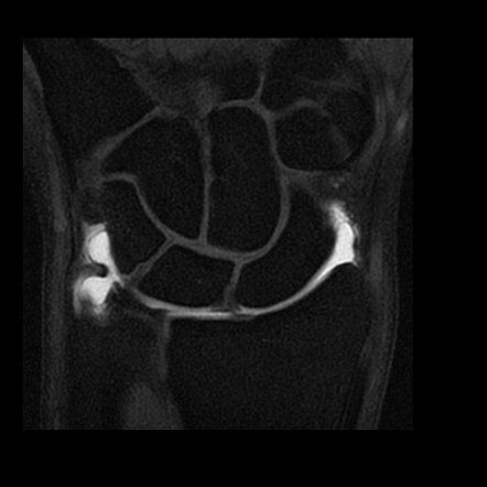
Arthrogram Mri Radiology Reference Article Radiopaedia Org

Learn More About An Arthrogram Of The Hip With This Patient Friendly Three Minute Video Magnetic Resonance Imaging Mri Radiologist

What You Need To Know About An Arthrogram Diagnostic Imaging Services
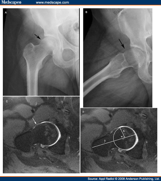
Diagnostic Imaging Of The Hip For Physical Therapists Physiopedia

Mri Arthrogram Hip Rule Out Labral Tear Cedars Sinai
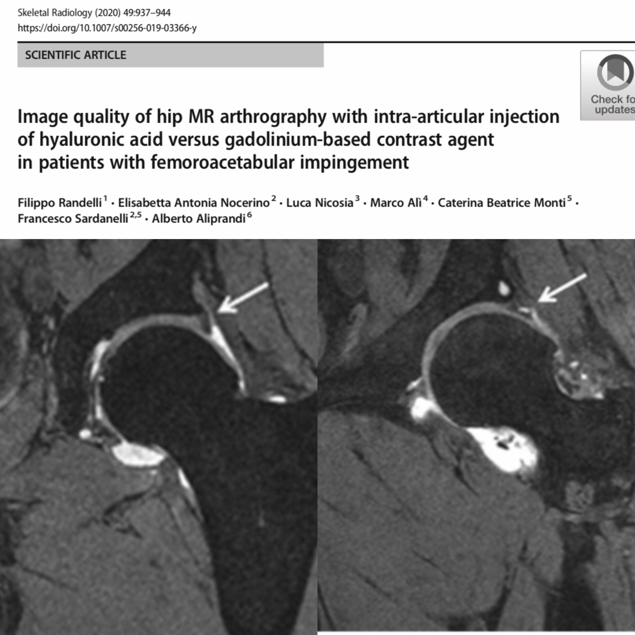
Skeletal Radiology Image Quality Of Hip Mr Arthrography With Intra Articular Injection Of Hyaluronic Acid Versus Gadolinium Based Contrast Agent In Patients With Femoroacetabular Impingement By Randelli Et Al T Co Jo5qw6kfk2

Mr Arthrogram For Joints Shoulder Elbow Wrist Hip Knee Ankle Mria S Suburban Imaging Twin Cities
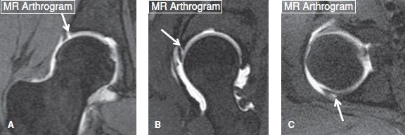
The Hip Musculoskeletal Key

Imaging Of A Painful Femoral Head Arthritis Research

Shoulder Mr Arthrogram Anatomy Of The Normal Glenohumeral Joint And Rotator Cuff

3t Mri As Good As Mr Arthrography For Planning Hip Surgery

Mri Arthrography Open Air Mri Of Cen La

Epos Trade
Journal Of Lancaster General Health Imaging Insights

Factors Influencing Discomfort During Anterior Ultrasound Guided Injection For Hip Arthrography Sciencedirect
Axial Cut Magnetic Resonance Mr Arthrogram Of The Left Hip Download Scientific Diagram

Arthrography And Injection Procedures Radiology Key

Mr Arthrogram T2 Weighted Fat Suppressed Sequence Of A Right Hip In Download Scientific Diagram

Mri Arthrogram Hip Rule Out Labral Tear Cedars Sinai
Coronal Section Mr Arthrography Of A Dysplastic Hip Th Open I

Hip Zona Orbicularis Mr Femoral Head Arthritis Research

Mri Shoulder Arthrogram Anatomy


